
skin surgery · dermatology · dermoscopy · Gdańsk
A mole, mole, mole, fibroma, wart or lump under the skin may be benign lesions, but sometimes they require diagnosis and removal with histopathological examination. The most important thing is to choose the method appropriate to the type of lesion - different treatment is required for a benign fibroma than for an oncologically suspicious mole.
Author: Editorial team of the Island of Friendly Medicine · Medical consultation: to be confirmed before publication · Publication: to be completed · Update: 08.07.2026
The article is educational in nature and does not replace a medical consultation. Any skin lesion that grows, bleeds, itches, changes color, hurts or looks unusual should be evaluated by a doctor.
The most important information at a glance
- Not every skin lesion is removed using the same method - the decision depends on its appearance, location, history and oncological suspicion.
- Suspicious lesions usually require surgical removal or surgical biopsy for histopathological examination.
- CO2 laser and cryotherapy can be used for selected benign lesions, but they are not an appropriate method for lesions suspected of melanoma.
- Dermoscopy helps assess moles and other skin lesions, but the final diagnosis of a surgically removed lesion is made by histopathology.
- Do not remove, burn or irritate moles or skin bumps yourself.
What skin lesions can be removed?
Patients most often come to the doctor with changes such as moles, moles, atheromas, fibromas, lipomas, warts, seborrheic keratosis, hemangiomas, skin cysts and subcutaneous nodules. Some of them are removed for health reasons, others due to irritation by clothing, bleeding, recurrent inflammation, pain or aesthetic reasons.
The most important question is not only "can it be removed?", but above all: "does the lesion require dermoscopy, surgical biopsy or planned excision with histopathological examination before removal?"
Diagnostics of skin lesions - from clinical assessment to histopathology
Diagnostics begins with an interview and viewing the lesion. The doctor asks, among other things: how long the lesion has existed, whether it is growing, bleeding, itching, painful, changing color, whether it has been irritated and whether there is a family history of skin cancer. It then assesses the location, size, color, boundaries, symmetry and structure of the lesion.
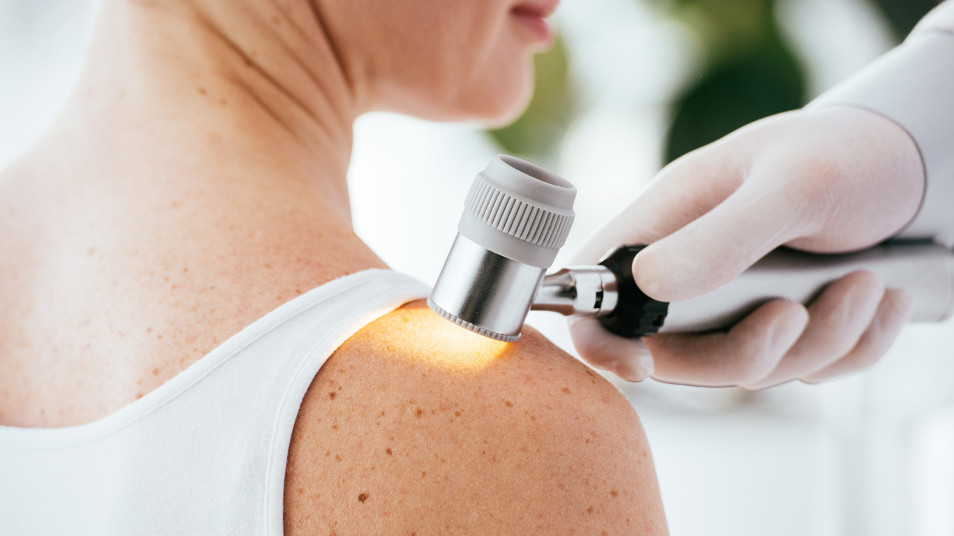
In the case of birthmarks and pigmented changes, the examination is important dermoscopy. This is a non-invasive magnified skin assessment that helps distinguish many benign lesions from those requiring further diagnostics. If a lesion is suspected of being cancerous, it should not usually be destroyed with a laser or cryotherapy - material for histopathological examination is needed.
In practice, this means that in case of suspicious lesions, the doctor may make suggestions surgical skin biopsy or surgical removal of the entire lesion and submitting it for histopathological examination.
Melanoma – when should a mole be a concern?
Melanoma is a skin cancer that may develop on the basis of an existing mole or appear as a new lesion. Early diagnosis is important, so it is worth knowing the ABCDE rule:
- A - asymmetry: one half of the change is different from the other.
- B - edges: the edges are uneven, jagged, blurred or irregular.
- C - color: the lesion has several colors or uneven coloration.
- D - Diameter: Lesions larger than approximately 6 mm are of particular concern, although smaller ones may also require evaluation.
- E - evolution: the lesion grows, changes shape, color, bleeds, itches, hurts, or looks different than before.
Not every asymmetric or dark lesion is melanoma, but any lesion that changes or looks unusual should be evaluated by a doctor. If melanoma is suspected, it is crucial to obtain material for histopathological examination.
Dermatologist or surgeon - who should evaluate and remove the lesion?
Dermatologists and surgeons often cooperate in the diagnosis of skin lesions. A dermatologist usually deals with skin assessment, dermoscopy, identification of pigmented lesions and qualification for further diagnostics. The surgeon excises the lesion, treats the wound, performs skinplasty, if necessary, and sends the material for histopathological examination.
The term "dermatosurgery" is commonly used for skin treatments performed in the field of dermatology and surgery. For the patient, the most important thing is not the nomenclature itself, but the proper qualification: oncologically suspicious lesions should be removed so that a reliable histopathological assessment and planning of further treatment is possible.
At the Friendly Medicine Island, diagnosis and treatment of skin lesions may include dermatological consultation, dermoscopy, surgical consultation, surgical skin biopsy and surgical removal of the lesion with histopathological examination - depending on the indications.
Methods of removing skin lesions
The method is selected by the doctor after assessing the lesion. The choice depends on whether the lesion is benign, suspicious, inflammatory, subcutaneous, pigmented, mechanically irritated, located in an aesthetically visible place or requiring histopathological examination.
1. Surgical excision of the skin lesion
Surgical excision involves the removal of the lesion under local anesthesia, usually with a tissue margin selected according to the indications. The wound is closed with stitches and the removed material may be sent for histopathological examination. This is especially important in the case of atypical, growing, bleeding, pigmented or oncologically suspicious lesions.
More information: surgical removal of skin lesions with histopathological examination in Gdańsk.
2. Surgical skin biopsy
Surgical skin biopsy is a diagnostic procedure. It involves taking a fragment of the lesion or excising the entire lesion in order to submit the material for histopathological examination. It may be needed when the image of the lesion is unclear or the doctor wants to confirm the diagnosis before further treatment.
More information: surgical skin biopsy in Gdańsk.
3. CO2 laser
The CO2 laser can be used for selected, benign skin lesions, e.g. some fibromas, warts or aesthetic changes, if the doctor does not see any signs of oncological suspicion. The advantage of the laser is precision, but a significant limitation is that the damaged tissue is usually not suitable for full histopathological evaluation.
Therefore, the laser should not be the first choice for lesions suspected of melanoma or other skin cancer.
4. Cryotherapy
Cryotherapy, i.e. freezing the lesion with liquid nitrogen, can be used for selected benign lesions, e.g. parts of viral warts or seborrheic keratosis. The treatment may cause burning, pain, blistering, discoloration or discoloration of the skin. Like laser, cryotherapy is not appropriate for lesions that require histopathological examination.
Why are suspicious lesions not removed with a laser?
If the lesion may be skin cancer, it is crucial to recognize it under a microscope. Laser or cryotherapy destroys tissue, which may prevent full histopathological evaluation. In the case of suspicious lesions, a safer path is usually surgical excision or surgical biopsy according to the doctor's qualifications.
This is especially important for pigmented lesions that change, bleed, itch, are asymmetric, have irregular edges or different colors.
What does surgical removal of a skin lesion look like?
The procedure is performed on an outpatient basis, most often under local anesthesia. The exact course depends on the type of lesion, its size, location and treatment plan.
- Consultation and qualification: the doctor assesses the lesion, indications for removal and possible method.
- Local anesthesia: the treatment area is anesthetized to reduce pain during the procedure.
- Excision of the lesion: the surgeon removes the lesion to the extent appropriate to the indications.
- Wound closure: the wound is closed with sutures, a dressing, and sometimes with a skinplasty technique.
- Histopathological examination: the removed tissue is subjected to microscopic evaluation, if indicated or consistent with the standard of a given procedure.
- Control and discussion of the result: the doctor provides recommendations, discusses the histopathology result and possible further treatment.
Convalescence after removal of a skin lesion
Healing time depends on the location, size of the lesion, type of procedure, skin tension, comorbidities and individual predispositions. After surgical removal of the lesion, the patient usually receives recommendations regarding dressing, washing the wound, limiting exercise, monitoring for symptoms of infection, and when to check-up or remove the stitches.
In the case of lesions on the face, neck, cleavage or other visible areas, the doctor may plan the cutting and dressing of the wound to reduce the risk of a visible scar. However, it is impossible to promise the absence of scars - each surgical intervention leaves a mark, the appearance of which depends on many factors.
When to report sooner?
Do not postpone consultation if the skin lesion:
- is growing rapidly or changing shape,
- is bleeding, oozing, forming a scab or ulcer,
- has multiple colors or uneven pigmentation,
- is asymmetric or has irregular edges,
- it itches, hurts or is constantly irritated,
- appeared suddenly and looks different from other moles,
- recurs after previous removal,
- is located in a place that is constantly damaged by clothing, belt, bra, razor or shoes.
Prevention of melanoma and other skin cancers
Prevention does not only involve removing lesions. Observation of the skin, protection against UV radiation and regular inspection of moles are of great importance, especially in people with fair skin, numerous moles, previous sunburn, use of solariums or a family history of skin cancer.
- Monitor your skin and look out for new or changing moles.
- Use sun protection and avoid sunburn.
- Do not use the solarium.
- Do not remove the lesions yourself using corrosive preparations or home methods.
- Perform dermoscopy as recommended by your doctor, especially if you have many moles or risk factors.
Removal of skin lesions on the Island of Friendly Medicine in Gdańsk
At the Friendly Medicine Island, patients can benefit from diagnosis and treatment of skin lesions depending on the indications: dermatological consultation, dermoscopy, surgical consultation, surgical skin biopsy and surgical removal of the lesion with histopathological examination.
Surgical procedures for skin lesions are performed by doctors specializing in skin surgery and small outpatient procedures, including: Piotr Rak, MD, PhD, Iwona Chruścicka, MD, PhD and Kamil Smok, PhD. The current scope of services and availability of doctors should be confirmed during registration.
In dermatological diagnosis, consultations and dermoscopy with dermatologists are helpful, including: of Dr. Andżelika Schwann-Majewska and of Dr. Katarzyna Przekazińska-Boager. Before publication, it is worth confirming current profiles and the scope of consultations.
Removal of lesions with histopathology Dermoscopic examination
FAQ - removal of skin lesions
Does every skin lesion need to be removed?
No. Some benign changes are enough to be observed. Removal may be indicated when the lesion grows, bleeds, hurts, irritates, recurs, looks unusual, or interferes with the patient functionally or aesthetically.
Can any mole be removed with a laser?
No. Pigmented lesions and oncologically suspicious lesions should not be destroyed with a laser if histopathological evaluation is needed. In such situations, the doctor usually considers surgical excision or surgical biopsy.
Is histopathological examination necessary after removal of the lesion?
Histopathological examination is particularly important in the case of atypical, growing, bleeding, pigmented, recurrent or suspicious lesions. It allows you to evaluate the removed tissue under a microscope.
Does it hurt to remove a skin lesion?
The surgical procedure is most often performed under local anesthesia. The patient may feel a sting, pressure or pull when the anesthesia is administered, but the purpose of the anesthesia is to reduce pain during the procedure.
Will there be a scar after the lesion is removed?
After surgical excision, a scar may always remain. Its appearance depends on the location, size of the lesion, suturing technique, wound care, individual healing and predisposition to scarring.
When should I undergo dermoscopy?
It is worth undergoing a dermoscopy if you have new or changing moles, numerous birthmarks, fair skin, a history of sunburn, a family history of melanoma, or when your doctor recommends monitoring the changes.
Can I remove a wart, mole or mole myself?
It is not worth removing the changes yourself. Home methods may result in burns, infection, scarring, incomplete removal of the lesion and delay in diagnosis if the lesion turns out to be suspicious.
Sources
- American Academy of Dermatology, ABCDEs of melanoma: https://www.aad.org/public/diseases/skin-cancer/find/at-risk/abcdes
- NICE, Melanoma: assessment and management: https://www.nice.org.uk/guidance/ng14
- NICE, Melanoma recommendations: https://www.nice.org.uk/guidance/ng14/chapter/recommendations
- NCBI Bookshelf, Guideline-based approach to cutaneous malignant melanoma: https://www.ncbi.nlm.nih.gov/books/NBK572149/
- Wyspa Medycyny Przyjaznej, surgical removal of skin lesions with histopathological examination: https://www.wyspamedycynyprzyjaznej.pl/pl/usuniecie-powiedz-skornych-z-badaniem-histopatologicznem-gdansk
- Wyspa Medycyny Przyjaznej, dermoscopy: https://www.wyspamedycynyPrzyjaznej.pl/pl/dermoskopia-badanie-mistrz-skornych
- Island of Friendly Medicine, surgical skin biopsy: https://www.wyspamedycynyPrzyjaznej.pl/pl/biopsja-zdrowie-skory-gdansk