
A biopsy is a key diagnostic test that allows doctors to precisely assess the nature of an abnormal lesion in the body. When diseases of the breast, skin, salivary glands or soft tissues are suspected, two methods are most commonly used: fine-needle biopsy (FNAB — fine-needle aspiration biopsy) and core needle biopsy (CNB). How do they differ, when is each method chosen, and why are they so important in oncological diagnostics? In this article, we explain it step by step. At our clinic, diagnosis and treatment of breast diseases and skin lesions are provided, among others, by Dr Iwona Chruścicka, MD, PhD, an oncological surgeon with many years of experience.
Fine-needle biopsy is a minimally invasive method that involves collecting cellular material using a thin needle. It is quick, usually performed under local anaesthesia, and allows cytological — cellular — assessment of the collected material. FNAB is particularly useful in:
The advantages of FNAB are simplicity, minimal discomfort and low cost. Its limitation is the small amount of material collected, which means that FNAB does not always provide a complete picture and sometimes needs to be supplemented with a more precise method.
Core needle biopsy involves collecting a larger tissue fragment with a thicker needle, often under ultrasound or mammography guidance. This makes it possible to perform a histopathological examination, which provides significantly more information about the nature of the lesion. CNB is used primarily in:
CNB is slightly more invasive than FNAB, but its results are more accurate — that is why in many cases it is irreplaceable.
| Feature | Fine-needle biopsy (FNAB) | Core needle biopsy (CNB) |
|---|---|---|
| Material collected | cells — cytological examination | tissue fragment — histopathological examination |
| Precision | initial assessment | higher — assessment of tissue structure |
| Invasiveness | lower, short recovery | slightly higher |
| Role | initial / screening test | confirmatory test / complex lesions |
“It is not about which method is better, but which one is appropriate for a given lesion. We often start with fine-needle biopsy, and when we need a full histopathological picture, we perform core needle biopsy. The method is selected after examination and imaging.”
— Dr Iwona Chruścicka, MD, PhD, oncological surgeon
Do you need a biopsy?
See the details of specific tests available at our clinic in Gdańsk and book an appointment:
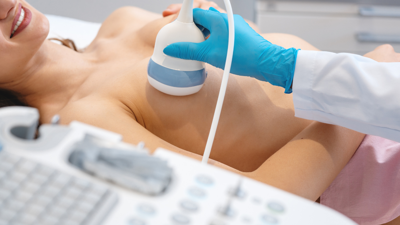
Fine-needle breast biopsy → Core needle breast biopsy → Surgical skin biopsy →The choice depends on the location and nature of the lesion, as well as on the results of imaging tests. The correct order is important: imaging diagnostics, such as ultrasound or mammography, are performed first, and only then is a biopsy carried out — often under ultrasound guidance, which allows the needle to be placed precisely in the lesion. The final decision on the type of biopsy is made by the doctor after examination.
Both methods play a key role in oncology. In breast cancer, the result of core needle biopsy allows doctors to determine the type of tumour, its aggressiveness and sensitivity to hormonal or targeted treatment. In melanoma diagnostics, biopsy allows the depth of invasion to be assessed, which influences further management. Similarly, in lesions of the salivary glands or soft tissues, biopsy provides information necessary to plan therapy.
If you are specifically interested in a breast examination — what it looks like step by step, whether it hurts and how to prepare — we have described it in detail in a separate guide on breast biopsy. Diagnostics are carried out by a team of oncological surgeons and radiologists, including Dr Iwona Chruścicka, MD, PhD, with many years of experience in oncological breast surgery.
Do you have a concerning lesion? Do not delay diagnostics.
Book an appointment at Wyspa Medycyny Przyjaznej in Gdańsk — we will choose the right method and quickly establish the diagnosis.
Book an appointment →Fine-needle biopsy and core needle biopsy are two complementary diagnostic methods. FNAB is faster and less invasive — it works well as an initial test. CNB provides a fuller histopathological picture and is the gold standard for assessing suspicious breast lesions. The choice is made by the doctor after imaging examination. If you notice a concerning lesion, do not delay — early diagnostics is the best protection for your health.
This article is for informational purposes only and does not replace a medical consultation. The choice of diagnostic method and interpretation of results are determined by the doctor.