
Informational article. If you are looking for a mole examination, dermoscopy or a dermatology consultation in Gdańsk, go directly to the examination or consultation page — the link is provided below.
Melanoma is a malignant skin cancer that may develop from an existing mole or appear as a new lesion. Early recognition is crucial: a mole that grows rapidly, changes colour, has irregular borders, bleeds, itches or looks different from the others requires dermatological assessment and often dermoscopy.
This article explains what melanoma can be mistaken for, how to use the ABCDE rule, when not to delay a consultation and how a suspicious skin lesion is diagnosed. At Wyspa Medycyny Przyjaznej in Gdańsk, diagnostics of skin lesions can begin with dermoscopy or a dermatology consultation.
Author: Wyspa Medycyny Przyjaznej Editorial Team · Medical consultation: to be completed by a dermatologist or surgical oncologist · Publication: to be completed · Updated: 7 July 2026
Do you have a new or changing mole?
Do not assess it only “by eye”. Dermoscopy allows the doctor to examine the structure of the lesion more closely and decide whether observation, follow-up or excision with histopathological examination is needed.
Book dermoscopy of skin lesions →Key information at a glance
| Question | Answer |
|---|---|
| What should be concerning? | asymmetry, uneven borders, multiple colours, rapid growth, bleeding, ulceration, itching or a change in the appearance of a mole |
| What is ABCDE? | a simple rule for assessing moles: asymmetry, borders, colour, diameter and evolution |
| Is every suspicious mole melanoma? | no, but every suspicious or changing lesion is worth examining |
| What does diagnosis involve? | skin examination, dermoscopy and, if melanoma is suspected, excision of the lesion with histopathological examination |
| Where should you start? | dermoscopy or a dermatology consultation |
What is melanoma?
Melanoma is a malignant tumour arising from melanocytes, the cells that produce skin pigment. It may occur on the skin, but also in less obvious locations, such as beneath a nail, on mucous membranes or within the eye. Most often, the patient notices a new mole or a change in the appearance of an existing one.
Melanoma is not always very dark and does not always look “typical”. It may be brown, black, red, pink, bluish or sometimes only lightly pigmented. When assessing moles, colour is therefore not the only relevant feature; change over time, asymmetry, irregular borders and difference from other lesions are also important.
According to data from the Polish National Cancer Registry, 4,028 new cases of cutaneous melanoma were registered in Poland in 2022. This number shows that prevention and early diagnosis of moles have real health significance.
“The patient does not have to decide independently whether a mole is dangerous. Their task is to notice a change and report it for assessment. Dermoscopy provides detailed diagnostics, and histopathological examination is used when necessary.”
— expert comment, subject to approval by a WMP doctor
The ABCDE rule — how to recognise a suspicious mole
The ABCDE rule helps patients assess whether a mole requires consultation. It does not replace a medical examination, but it helps identify changes that should not be ignored.
| Letter | Meaning | What should be concerning? |
|---|---|---|
| A | asymmetry | one half of the lesion looks different from the other |
| B | borders | the borders are uneven, ragged, blurred or irregular |
| C | colour | several colours are visible within one lesion: brown, black, red, grey, blue or areas of depigmentation |
| D | diameter | the lesion is larger than 6 mm or is growing, although melanoma may also be smaller |
| E | evolution | the mole changes size, shape or colour, becomes raised, bleeds, itches or hurts |
The most important letter is E, meaning evolution. A lesion that changes should be examined even if it does not meet all the other criteria.
The “ugly duckling” sign — when one mole looks different from the rest
In addition to the ABCDE rule, it is worth paying attention to the so-called ugly duckling sign. This refers to a lesion that clearly differs from the patient’s other moles. It may be darker, lighter, larger, more irregular or simply “different”.
If most moles look similar and one lesion deviates from this pattern, it is worth showing it to a dermatologist. This is particularly important in people with numerous moles, fair skin, a history of sunburn or a family history of melanoma.
What can melanoma be mistaken for?
Melanoma can resemble many benign skin lesions. Self-assessment based on internet photographs can therefore be misleading. On the other hand, many lesions that look worrying to the patient turn out to be benign after dermoscopy.
Melanoma may be confused with, among other things:
- a benign melanocytic naevus — an ordinary mole that is stable and regular,
- seborrhoeic keratosis — a rough, often brown lesion that may look as though it is “stuck on” the skin,
- lentigo — a flat pigmented lesion associated, among other factors, with sun exposure and age,
- angioma — a red or bluish-red vascular lesion,
- a scab after an injury — especially if the patient does not remember damaging the skin,
- a subungual haematoma — which may resemble subungual melanoma,
- an inflammatory or infectious lesion — when redness, discharge or ulceration is present.
If a lesion bleeds, does not heal, grows, returns after a scab falls off or appeared without a clear cause, it requires medical assessment.
When should you see a dermatologist or surgeon?
Not every skin lesion requires urgent treatment, but some symptoms should not be postponed. It is better to examine a lesion early than to observe it for months without knowing what it is.
Book a dermatology consultation or dermoscopy if you notice:
- a new mole that is growing rapidly,
- a change in the colour, shape or size of an existing mole,
- irregular borders or asymmetry,
- several colours within one lesion,
- bleeding, ulceration, discharge or crusting without injury,
- itching, pain, burning or tenderness of a mole,
- a dark streak under a nail that is getting larger,
- a lesion that looks different from all the others.
If the doctor considers the lesion suspicious, surgical excision and histopathological examination may be recommended. Suspicious lesions should not be removed with a laser, electrocoagulation or another tissue-destructive method that prevents histopathological assessment when cancer is suspected.
Could this be melanoma?
| Symptom | What might it mean? | What should you do? |
|---|---|---|
| a mole changes colour or shape | a lesion requiring dermoscopic assessment | book dermoscopy |
| bleeding or ulceration without injury | a warning sign | do not delay consultation |
| a lesion looks different from the other moles | the “ugly duckling” sign | show it to a doctor |
| a dark streak beneath a nail | a haematoma or pigmentation, but sometimes subungual melanoma | dermatological assessment, especially if the streak is enlarging |
| a scab that falls off and returns | a non-healing lesion | consultation and a decision about further diagnostics |
What does dermoscopy involve?
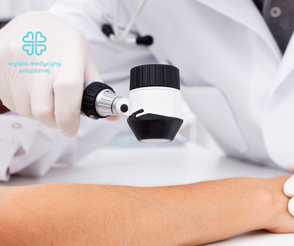
Dermoscopy is a non-invasive magnified examination of skin lesions. The doctor assesses the structure of the mole, pigment, blood vessels, symmetry and other features that are difficult to see with the naked eye. The examination is painless and usually brief.
Dermoscopy helps determine whether a lesion can be observed, whether follow-up after a specified period is needed, or whether it should be removed and submitted for histopathological examination. This is important because a definitive diagnosis of melanoma is made on the basis of histopathological assessment of the excised lesion.
What happens if the doctor suspects melanoma?
If a lesion looks suspicious, the doctor may recommend surgical excision of the entire lesion with an appropriate margin and submission of the tissue for histopathological examination. This examination determines whether the lesion is benign or malignant and, if it is melanoma, identifies features important for further treatment.
If melanoma is confirmed, further management depends on the histopathology result, depth of invasion, margins, location and any risk features. The patient may require wider excision, lymph-node diagnostics or treatment at an oncology centre. The scope of treatment is determined by the doctor or a multidisciplinary team.
If the doctor recommends removal, more information about the procedure is available here: removal of a skin lesion with histopathological examination.
Can melanoma be removed with a laser?
A suspicious pigmented lesion should not be destroyed with a laser, frozen or removed using a method that does not permit full histopathological examination. If melanoma is suspected, preserving tissue for microscopic assessment is crucial.
Laser treatment or other tissue-destructive methods may be used for selected benign lesions, but only after medical assessment. In the case of suspicious moles, oncological diagnostics take priority over the cosmetic result.
Who is at higher risk of melanoma?
Melanoma can occur in anyone, but risk is higher in people with certain factors. Awareness of risk helps plan skin self-examination and regular dermatological checks.
Risk factors include:
- fair skin, fair or red hair and a tendency to sunburn,
- numerous moles or atypical moles,
- a previous melanoma,
- a family history of melanoma,
- intense exposure to ultraviolet radiation,
- sunburn, particularly during childhood,
- use of tanning beds,
- reduced immunity, for example after transplantation or during immunosuppressive treatment.
How should you check your skin yourself?
Skin self-examination does not replace dermoscopy, but it helps identify changes that appear between visits. It is worth examining the skin in good light, preferably once a month, using a mirror or asking a close person to check the back and scalp.
During self-examination, check:
- the face, ears, neck and scalp,
- the chest, abdomen and back,
- the upper arms, forearms, hands and spaces between the fingers,
- the buttocks, groin, legs and feet,
- the soles of the feet and areas around the nails,
- moles that look different from the others.
Photographing lesions that cause concern may be helpful. A photograph does not replace dermoscopy, but it can make it easier to notice whether a mole is truly changing.
Melanoma prevention — how can risk be reduced?
The most important element of prevention is reducing excessive exposure to ultraviolet radiation. This does not mean avoiding sunlight completely, but protecting the skin sensibly, particularly during the hours of strongest sunlight.
Helpful preventive measures include:
- avoiding sunburn,
- using high-protection sunscreen and reapplying it regularly,
- wearing a hat, sunglasses and protective clothing,
- avoiding tanning beds,
- protecting children’s skin,
- self-examination of moles,
- dermoscopy in people with numerous moles or risk factors.
Who should you see at Wyspa Medycyny Przyjaznej?
At Wyspa Medycyny Przyjaznej in Gdańsk, a patient with a suspicious mole, new skin lesion, bleeding growth or a lesion requiring dermoscopy may begin diagnostics with a dermatology consultation. If the lesion requires excision, the doctor may refer the patient to a surgeon.
surgery and surgical oncology — excision of skin lesions for histopathological examination
More about the doctor
Key information at a glance
- Melanoma may develop from an existing mole or appear as a new lesion.
- The ABCDE rule helps identify moles requiring assessment.
- A lesion that grows, bleeds, itches, hurts or looks different from the others requires medical assessment.
- Dermoscopy is a non-invasive examination that helps assess the structure of a mole.
- If melanoma is suspected, excision of the lesion and histopathological examination are crucial.
- Suspicious moles should not be destroyed using methods that do not allow histopathology.
- UV protection and regular skin checks reduce the risk of late diagnosis.
Frequently asked questions
Is melanoma always black?
No. Melanoma may be black or brown, but it may also be red, pink, grey, bluish or only lightly pigmented. Colour is therefore not the only important feature; change over time, asymmetry, borders and difference from other moles also matter.
Does every large mole need to be removed?
Not every large mole requires removal. The decision depends on clinical appearance, dermoscopy, changes over time and the patient’s risk factors. Sometimes observation or follow-up is sufficient, while in other cases the doctor recommends excision and histopathological examination.
When does a mole require prompt dermoscopy?
Dermoscopy should be performed promptly if a mole grows, changes colour or shape, bleeds, itches, hurts, has uneven borders, ulcerates or looks different from the other moles. A new dark streak beneath a nail is also concerning.
Is dermoscopy painful?
No. Dermoscopy is non-invasive and painless. The doctor examines the lesion using a specialised magnifying device. The examination does not require cutting the skin and helps determine whether the lesion can be observed, followed up or should be excised.
Can melanoma be diagnosed without excision?
Dermoscopy can strongly suggest whether a lesion is benign or suspicious, but a definitive diagnosis of melanoma is made by histopathological examination. Suspicious lesions therefore usually require surgical excision and microscopic assessment.
Can a suspicious mole be removed with a laser?
A suspicious mole should not be destroyed with a laser or another method that prevents histopathological examination. If melanoma is suspected, the priority is to excise the lesion in a way that allows microscopic assessment.
Which doctor should assess a suspicious mole?
It is best to begin with a dermatologist and dermoscopy. If the lesion requires excision, the doctor may refer the patient to a surgeon who removes it and sends the tissue for histopathological examination.
Have you noticed a new, changing or bleeding mole?
Do not wait for further changes. Dermoscopy helps determine whether the mole is benign, requires follow-up or should be excised and examined histopathologically.
Book dermoscopy →Summary
Melanoma is a malignant skin cancer that may resemble an ordinary mole, seborrhoeic keratosis, lentigo, a subungual haematoma or a post-traumatic lesion. The most important warning signs are asymmetry, irregular borders, multiple colours, growth, bleeding, ulceration, itching, pain and a lesion that looks different from the other moles.
Early diagnostics are based on skin examination and dermoscopy. If a lesion is suspicious, the doctor may recommend surgical excision and histopathological examination. Suspicious moles should not be removed with tissue-destructive methods that prevent microscopic assessment.
If you have a mole that is changing, book dermoscopy in Gdańsk or a dermatology consultation.
You may also be interested in
Sources and medical context:
- American Academy of Dermatology — ABCDEs of melanoma
- DermNet — ABCDE signs of melanoma
- NICE NG14 — Melanoma: assessment and management
- ESMO Clinical Practice Guideline — Cutaneous melanoma
- Polish National Cancer Registry / Pacjent.gov.pl — melanoma epidemiology
- Wyspa Medycyny Przyjaznej — dermoscopy of skin lesions
This article is informational and does not replace a medical consultation. A new, growing, bleeding, ulcerated, itching, painful or changing mole requires individual dermatological assessment. The doctor decides on the diagnosis, need for excision, histopathological examination and further treatment.